Showing posts with label Critical Care. Show all posts

Wednesday, October 21, 2015

For the Sake of the Trach: Tracheostomy Basics & Complications in the ED

Clinical scenario: You’re working in the ED when you receive a pre-arrival page: 62 y M with trach in respiratory distress, unable to obtain sats, ETA 5 min.  As you are setting up suction in the resuscitation bay, EMS arrives with a pale, elderly male gasping for air.  What's your next move?

Review: Although (hopefully) not something we see in the emergency department every day, this is absolutely something we need to be comfortable stabilizing, if not definitively managing, on our own. In order to have a better understanding of tracheostomy complications, it’s important to understand some terminology and anatomy first.  

Differentiating tracheostomies from laryngectomies 
A critical piece of information to obtain in patients with a tracheostomy is to determine whether or not they have had a laryngectomy as well. A tracheostomy is simply an opening in the trachea created with an incision through the anterior neck. Some reasons for needing a tracheostomy include chronic mechanical ventilation, maxillofacial trauma, or upper airway obstruction such as from a mass. A laryngectomy—usually performed on patients with laryngeal cancer—is a complete removal of the larynx with separation of the airway from the mouth, nose, and esophagus. Since there is no connection between the mouth and the patient’s airway, laryngectomy patients can NEVER be orally
intubated. Outwardly, a laryngectomy patient looks like any other patient who has had a tracheostomy, so it is impossible to tell if a patient has had a laryngectomy simply by looking at his or her neck. While occluding the stoma of a patient who has only had a tracheostomy may not cause complete loss of the ability to ventilate (assuming that have at least a somewhat patent upper airway), occlusion of the stoma in a larygectomy patient will absolutely in all cases make it impossible for the patient to breathe, since the stoma is the only possible connection to the lungs in a laryngectomy patient. Despite being a “never” event, attempts at oral ventilation on a laryngectomy patient have been reported in the past [1].   If a patient with a total laryngectomy requires bagging and the trach tube is displaced, the stoma is the only way to ventilate them.  As an initial measure, an laryngeal-mask airway or neonatal mask can be applied to the stoma and used for bagging.


Tracheostomy basics 
To better understand tracheostomies in general, some basics are worth reviewing. Tracheostomies can be performed percutaneously at the bedside in the intensive care unit or surgically in the operating room. Various methods for a tracheostomy exist, but the neck incision is usually made midway between the cricoid cartilage and the sternal notch, well below where a cricothroidotomy is usually performed, and the trachea itself may be opened with a vertical or horizontal incision. A few safety features are sometimes integrated into the tracheostomy: stay sutures and Bjork flaps. Stay sutures are temporary sutures placed through 2-3 tracheal rings that allow for the trachea to be pulled back up to the skin should decannulation occur. This allows for visualization and easier reinsertion of the tracheostomy tube, thereby decreasing the risk of creating a false passage if the tracheostomy tube needs to be reinserted before the tract fully matures in about 7 days. Stay sutures are usually removed after 7 days, so patients presenting to the ED are not likely to have these. A Bjork flap is an upside-down U-shaped section of trachea that not only creates the tracheostomy, but the free edge of the flap is sutured to the skin of the neck, essentially creating a path for tracheostomy tube reinsertion and reduces the risk of creating a false passage when reinserting a tracheostomy tube prior to tract maturation [2]. 

Tracheostomy Tube Features 
Tracheostomy tube designs vary widely but most tracheostomy tubes have a number of parts in common. A few important details to know about every tube include the size of the tube, the brand or type, and whether or not the tube has a cuff. For example, when speaking with a consultant, one might say, “this patient has a size 6 cuffed Shiley.” The cuff is an important feature of many
tracheostomies as it allows for the airway to be sealed off, allowing for positive pressure ventilation and reducing the risk of aspiration. Deflating the cuff will allow the patient to breath through his or her mouth to some degree (assuming a patent upper airway), which serves as a backup for ventilation should the tube become occluded. Having the cuff down also allows for the patient to phonate when they occlude their tracheostomy tube, since air will be able to pass around the deflated cuff and tube through the vocal cords. Some tracheostomy tubes will have both an outer cannula and inner cannula, which allows for the inner cannula to be removed and cleaned or replaced without changing the entire tracheostomy tube. The downside to having an inner cannula, however, is that the effective inner diameter of the tube is decreased, so the patient may experience increased resistance to airflow.


Tracheostomy tube complications 
Setting up for the patient
Patients with tracheostomy tubes will on occasion present to our emergency department, and as the initial responders to these emergencies, it is important to be aware of the common or potentially life threatening complications associated with tracheostomy tubes. In the patient who is not rapidly decompensating, eliciting a brief history focused on the tracheostomy tube should be performed. Necessary details such as when the tracheostomy was placed, what size tube the patient uses, and why the tracheostomy was needed may be management altering pieces of information. When the patient arrives to the ED, supplies and equipment should be assembled in anticipation of potential worsening of the patient's complication. Personal protective equipment (face shield, gloves, fluid-resistant gown), suction catheters, Yankauer suction, replacement tracheostomy tubes (of the same and also one size smaller), tracheostomy tube ties, and a supplemental oxygen source should all be at the bedside ready for immediate use. Endotracheal tubes with intubating equipment (if the patient has a patent upper airway) should be readily available as well, if not at the bedside. 


Tracheostomy tube obstruction
Secretion buildup will often result in a narrowing of the effective tube diameter, commonly leading to increased resistance to flow and manifesting as respiratory distress in the patient. Inadequate suctioning, poor hydration, and decreased mobility are all risk factors for obstruction from secretions. The initial step in patients with a possible tube obstruction is to attempt passing a suction catheter through the tracheostomy tube. Instilling a few milliliters of sterile saline may help loosen secretions. If the suction catheter cannot be passed easily beyond a few centimeters or the length of the tube, the tube may either be obstructed or dislodged. In tubes with an inner cannula, the inner cannula should be removed and inspected or replaced, but if there is still resistance to passing a suction catheter, the tracheostomy tube is likely dislodged with the distal tip in the soft tissues of the neck and will need to be removed immediately and replaced [2]. 


Accidental decannulation
Although most patients will have a mature tracheostomy tract when they present to the ED, it is prudent to ask when the tracheostomy was placed. Tracheostomies that are less than 7 days old presenting with a decannulation of the tube should never be replaced blindly because of the risk of creating a false passage upon reinsertion. In a patient with a mature tract, he or she should be optimally positioned for reinsertion, preferably laying supine with a shoulder roll to extend the neck, which will help align the tissue planes and mimic the position by which the tracheostomy was originally created (likely supine on a operating table). Preoxygenating the patient oronasally or via the stoma will reduce the risk of oxygen desaturation should any difficulties arise during the procedure. Always use an obturator or introducer if available to avoid injuring the soft tissues with the end of the tracheostomy tube. Water-soluble lubricant or a lidocaine containing jelly should be applied to the tube. Holding the tube and obturator as one unit, the tube should be inserted with the tip initially pointed perpendicular to the stoma and then gently curved downward into the trachea following the bend of the tube. If the tube has been out of the stoma for more than several hours, the stoma may have begun to stenose and require dilation by an otolaryngologist prior to reinserting a tube. A chest x-ray should be performed to confirm placement. Alternatively, if available, a nasopharyngoscope or bronchoscope can be used to directly visualize the carina via the tracheostomy tube, which would guarantee proper tracheal placement.  For a video demonstration, see this you tube video.


Bleeding from the tracheostomy
Tracheostomy bleeds can be from a number of possible sources. Superficially, the skin underlying the flange of the tracheostomy tube should be checked, as malpositioning of the tube or patient may result in pressure ulceration. The tracheostomy tube may need to be removed to fully inspect the stoma and surrounding skin, and local bleeding can be controlled with pressure or topical silver nitrate. Often, granulation tissue, which are new growths of connective tissue and small blood vessels, can arise from the stoma site, or even within the trachea itself. Minor bleeding from around the stoma can similarly be treated with pressure or silver nitrate. Granulation tissue within the trachea is diagnosed by direct visualization with a nasopharyngoscope or bronchoscope, and needs to be definitively treated by ENT, usually by cauterization. Other potential sources of tracheostomy bleeding may come from the tube eroding into the thyroid vessels, thyroid gland, or tracheal wall. A tracheoinnominate fistula is perhaps the most feared complication of a tracheostomy tube and occurs when the tip of the cannula erodes through the anterior tracheal wall and into the innominate artery. This rare condition occurs in less than 1% of all patients with a tracheostomy tube but carries a mortality rate approaching 100% given the catastrophic bleeding into the airway. Approximately 75% of patients with a tracheoinnominate fistula will present within 3 to 4 weeks of tracheostomy tube placement, and some of these patients will have an initial "sentinel bleed" that may be relatively minor before developing massive hemorrhage [2,4]. Hemorrhage in these cases can be temporized by hyperinflating the cuff of the tracheostomy tube or endotracheal tube placed through the stoma as an attempt to tamponade the bleeding. These patients will need emergent thoracic and ENT consultation. Endovascular embolization of the innominate artery may be another option in these patients and has been demonstrated to be successful in a few case reports [5]. 


Cardiac arrest
Suppose a patient with a tracheostomy is brought into the emergency department with CPR in progress. Provided that the patient's stoma remains patent, a small cuffed endotracheal tube (e.g. a 6.0 tube) can be inserted through the stoma to ventilate a tracheostomy patient in this code scenario. Intubation of the stoma is not only is much faster than attempting oral intubation, but also avoids the potential attempt at oral intubation on a laryngectomy patient (which, again, should never occur) if his or her medical history is unknown. A laryngeal mask airway (LMA) can be placed over the stoma to ventilate if an endotracheal tube is not readily available, but the patient's mouth and nose should be covered if upper airway patency is unknown. Alternatively, should the stoma be stenosed (for example, if the tracheostomy tube has been out for hours) and the patient is known to have a patent upper airway, the stoma can be occluded and the patient can be ventilated with bag-valve-mask via the mouth and nose. 


Take Home Points 
Tracheostomy complications can quickly become life-threatening, and knowing some basic concepts about tracheostomies can allow us to better respond to and take care of patients with these complications. As with any patient, getting an adequate history should be the first step, and in particular, knowing if the patient has had a laryngectomy can prevent the “never event” of an orotracheal intubation attempt. Before performing any interventions on a patient where there is time to set up (i.e. on a relatively stable patient), one should gather appropriate equipment such as personal protective gear, extra tubes, and suction. Finally, consider the potential for a tracheo-innominate fistula in a bleeding tracheostomy patient given the extremely high associated mortality. 

Submitted by Phil Chan, PGY-3
Faculty Reviewed by jason wagner  (@TheTechDoc)
Everyday EBM Editor: Maia Dorsett (PGY-4, @maiadorsett) 

References

[1] El-Sayed IH, et al. Identifying and Improving Knowledge Deficits of Emergency Airway Management of Tracheotomy and Laryngectomy Patients: A Pilot Patient Safety Initiative. Int J Otolaryngology. 2010;2010:1-7.
[2] Morris LL, et al. Tracheostomy Care and Complications in the Intensive Care Unit. Crit Care Nursing. 2013;33(5):18-30.
[3] D. Doyle J, Scales DC. Tracheostomy. In: Hall JB, Schmidt GA, Kress JP. eds. Principles of Critical Care, 4e. New York, NY: McGraw-Hill; 2015. http://accessmedicine.mhmedical.com/content.aspx?bookid=1340&Sectionid=80032214. Accessed September 24, 2015.
[4] Epstein SK. Late Complications of Tracheostomy. Respir Care 2005;50(4):542-549.
[5] Hamaguchi S, Nakajima Y. J Vasc Surg. 2012;55:545-547

Wednesday, April 29, 2015

A Balancing Act

It’s another busy day in the ED when an elderly female comes in from triage with fever, cough, and new oxygen requirement. Even before the patient comes back you are concerned for pneumonia with sepsis. The patient is tachycardic and hypotensive with a shock index greater than one. You institute early antibiotics and fluids and systematically begin to aggressively resuscitate her. The patient requires nearly four liters of normal saline before her blood pressure stabilizes. Your attending suggests that your liberal use of normal saline will induce a hyperchloremic metabolic acidosis, and perhaps you should have used lower chloride containing fluid, like lactated ringers. You perform a brief literature review on the topic of balanced resuscitation using lower chloride containing fluids.

Literature Review:
Strong Ion Difference (Kishen et al)
The main difference between normal saline and balanced fluids, such as lactated ringers, is the strong ions difference (SID), that is, the difference between cations (e.g. Na+) and anions (e.g. Cl-).  Normal saline has a SID of zero (equal parts Na+ and Cl-) where as Lactated ringers has a SID of 28, which is due to the additional cations such as Ca+, K+, and lower anion (Cl-) content.  Importantly, normal plasma SID content ranges from 38-44mmol/L, therefore balanced fluids more closely approximates physiologic SID.  As the SID becomes narrower, as is the case with significant normal saline administration, a non-gap metabolic acidosis develops. [1]

The use of normal saline in large volumes has been shown to produce a reliable drop in serum pH as demonstrated by Scheinraber et al, in a study among patients undergoing elective surgery. [2] However, the development of a hyperchloremic acidosis is of unclear clinical significance. Early animal models in dog kidneys demonstrated that compared to non-chloride fluids, chloride containing solution led to renal vasoconstriction and decline in glomerular filtration rate. Similarly a randomized, double blind crossover study in healthy humans demonstrated a significant reduction in renal blood flow and renal tissue perfusion, after the administration of two liters of normal saline compared to low chloride (98 mEq/L) Plasma-Lyte solution. [3] However, the effect of isotonic saline in acutely ill patients is still not as clear. A prospective cohort study among 175 ICU patients demonstrated that higher chloride levels (109.4 vs 115.1mEq) was an independent factor for increased mortality, although a limitation of this study was they could not distinguish the cause of hyperchloremia (iatrogenic, renal dysfunction, or endogenous hyperchloremia) [4]
Traditional and 'balanced' fluid content (crashingpatient.com)
A large retrospective cohort study of critically ill adults with vasopressor dependent sepsis showed lower in-hospital mortality in patients who received balanced (lower chloride) fluids versus isotonic saline, 19.6% versus 22.8% (RR 0.86; 95% CI,0.78-0.94). A limitation of this study was that patients receiving balanced solutions were younger, less likely to have chronic heart and renal failure, and more likely to receive steroids, colloids and invasive monitoring. [5] A 2014 retrospective study in 109,836 patients that met SIRS criteria and received crystalloid fluid resuscitation, showed that low-chloride loads were associated with lower in-hospital mortality. This mortality difference remained even after adjustment for severity of illness and total fluid volume administered. [6]

Similarly, a before and after study by Yunos et al involving 1644 ICU patients, reported the use of chloride-restricted fluids was associated with lower serum creatinine and decreased rates of renal replacement therapy (6 vs 10%) compared to controls. Like the study by Shaw et al, the difference was independent of severity of illness or total fluid volume administered. However, as mentioned by the authors, determining which component of lower-chloride fluid may have led to the observed effect is difficult, as there was simultaneous administration of lower sodium content, as well as increase in the administration of acetate, lactate, and gluconate. Importantly, this study showed no difference in mortality. [7][8]

Take home points: Administration of large volume of isotonic saline is associated with a metabolic acidosis. Animal models have demonstrated decreased renal perfusion with chloride containing fluids. Several retrospective studies indicate that chloride is an independent risk factor for mortality in acutely ill patients. More and more literature in humans seems to indicate that a ‘balanced resuscitation’ may decrease morbidity, and possibly mortality, in patients receiving large volumes of crystalloids as part of their resuscitation.  A single nonrandomized study demonstrated a correlation between low chloride fluids and decreased use of renal replacement therapy. Blinded, randomized, prospective studies are needed to further elucidate this observed effect.

Expert Commentary:

Dr. Schwarz, an Assistant Professor here at Wash U, and both an Emergency Physician and Toxicologist has provided some of his own thoughts on the topic. 

First, I’d like to thank Louis for picking a great topic and generating discussion about a very important subject.  I initially became interested in this topic a few years ago.  Originally, I was much more interested in the mechanism by which normal saline (NS) caused a non-anion gap metabolic acidosis, and that’s when I learned about the strong ion difference and a ‘balanced resuscitation.’  As a full disclosure while I found the pathophysiology really interesting, I initially didn’t think it had much clinical relevance.  However as more investigators have studied this, I’ve come to believe that my initial impressions were incorrect and changed my practice.

 The last time I reviewed the literature, I didn’t see a randomized, controlled trial comparing resuscitation with NS and lactated ringers in the ED.  However I do believe that there are studies out there that are applicable to the ED.  A retrospective study compared patients undergoing elective or emergent general surgery that received either NS or a ‘balanced fluid.’1  Unadjusted mortality and the number of patients developing major complications were higher in the group that received NS; after adjusting with propensity scoring, the mortality was no longer significantly different between the 2 groups.  However, patients that received NS were 4.8 times more likely to require dialysis. In a meta-analysis of patients with sepsis, patients that received a ‘balanced resuscitation’ had a lower mortality than patients receiving NS.2  The trend, however, was not significant.

In a promise to keep this short, I won’t review all the other literature that has been published on this topic and kept the discussion on the 2 articles that I did include short.  I’ll also concede that the literature is not perfect, and as I mentioned earlier, I’m also still waiting for that perfect ED-based study to be completed.  However the cost of NS or a ‘balanced solution’ such as lactated ringers is nearly equivalent.  I’m also not aware of significant complications from administering lactated ringers in most patients. So when the risks, costs, and benefits of implementing a ‘balanced resuscitation’ verses a standard resuscitation with NS are viewed together, I think there is enough evidence to consider changing your resuscitation strategy.

Now like many EDs, lactated ringers is not kept in our department.  It is on shortage but so is NS.  Neither of those are reasons not to use it.  So what do I do? Since I haven’t been able to convince pharmacy to keep lactated ringers in the ED yet, I do my best to guess early on which patients are going to need large-volume resuscitations.  If I think they are going to likely need more than 2-3 liters of fluid, I order additional lactated ringers from the pharmacy when I place their initial orders. In an hour after the patient has received the first few liters of NS, the lactated ringers should be there from the pharmacy.  If they need further resuscitation I can use it or return if they no longer need it.  For those that are interested to read more about this topic, I’d direct you to the upcoming May 2015 edition of Emergency Physicians Monthly. From my understanding, it’s brilliantly written! (Sorry for my shameless plug)


Jamtgaard References:
 [1] Kishen R, Honoré PM, Jacobs R, et al. Facing acid–base disorders in the third millennium – the Stewart approach revisited. International Journal of Nephrology and Renovascular Disease. 2014;7:209-217. doi:10.2147/IJNRD.S62126.
[2] Scheingraber et al. Rapid Saline infusions produces hyperchloremic acidosis in patients undergoing gynecologic surgery. Anesthesiology 1999;90;1265
[3] Chowdhury A et al. .  A randomized, controlled, double-blind crossover study on the effects of 2-L infusions of 0.9% saline and plasma-lyte® 148 on renal blood flow velocity and renal cortical tissue perfusion in healthy volunteers.  Ann Surg. 2012;256(1):18-24
 [4] Boniatti MM et al.  Is hyperchloremia associated with mortality in critically ill patients? A prospective cohort study. J Crit Care. 2011;26:175–179. doi: 10.1016/j.jcrc.2010.04.013
[5] Raghunathan K, Shaw A, Nathanson B et al. Association between the choice of IV crystalloid and in-hospital mortality among critically ill adults with sepsis*. Crit Care Med 2014; 42: 1585–91
[6] Shaw A et al.  Association between intravenous chloride load during resuscitation and in-hospital mortality among patients with SIRS. Intensive Care Medicine. 2014;40(12):1897-1905. doi:10.1007/s00134-014-3505-3.
[7] Waikar SS, Saving the Kidneys by Sparing Intravenous Chloride?.JAMA. 2012;308(15):1583-1585. doi:10.1001/jama.2012.14076.
[8] Yunos N et al. Association between a chloride-liberal vs chloride restrictive intravenous fluid administration strategy and kidney injury in critically ill adults. JAMA 2012; 308: 1566– 72.

Schwarz References
1. Shaw et al.  Major Complications, Mortality, and Resource Utilization after Open Abdominal Surgery: 0.9% saline compared to Plasma-Lyte. Ann Surg 2012;255:821-829.
2. Rochwerg et al. Fluid Resuscitation in Sepsis. A Systematic Review and Network Meta Analysis. Ann Intern Med 2014;161:347-355.


Submitted by Louis Jamtgaard +Louis Jamtgaard , PGY-3
Faculty Reviewed by Evan Schwarz @TheSchwarziee 

Wednesday, April 15, 2015

Does cardiac standstill on bedside echo equal 100% mortality?

You’re in the midst of catching up on notes during a hectic overnight shift when out of the corner of your eye you see a stretcher zoom into the trauma bay – with an EMT leaning over the side performing chest compressions. As the team gathers, the paramedics give report. The patient is a middle-aged male, no known past medical history, who was acting normally about half an hour ago when he suddenly collapsed in front of his family. They started CPR within a couple minutes of the patient collapsing, and called EMS. The paramedics continued CPR, placed a supraglottic airway, and placed the patient on the monitor. He has had a slow, organized rhythm without pulse throughout the arrest. He has received several doses of epinephrine without response. The patient has been pulseless for a little over half an hour by the time he arrives. The ED crew takes over CPR, IV access is obtained, and the patient switched over to the ER monitors, which show a slow, wide-complex, relatively disorganized rhythm. The patient shows no signs of life. Your attending physician calls for the ultrasound, and calls out to the team that if the bedside echo shows cardiac standstill, you will consider terminating further resuscitative efforts.

Clinical Question:

Does cardiac standstill on bedside echo universally predict mortality in OHCA?

Tuesday, April 7, 2015

Needle that belly!

An infant female with no significant history presents to your trauma bay after reported accidental blunt trauma to the abdomen, the patient arrives from a referral hospital where plain films demonstrated free air. On arrival the patient show signs of hemodynamic instability and an elevated lactate. The patient was decompressed with "needle peritoneumostomy" prior to going to the OR for exploration. 

Clinical Question:

Can “tension pneumoperitoneum” cause hemodynamic instability?

Literature Review:

The presence of "free air" in the peritoneum is often diagnostically significant; however, the gas itself is rarely of clinical importance. An exception to this rule is in the case of a tension pneumoperitoneum. Tension pneumoperitoneum (TPP), also known as hyperacute abdominal
Example of pneumoperitoneum & football sign
compartment syndrome [1], or abdominal tamponade [2], is a rare, but potentially deadly event. Similar to tension pneumothorax, the underlying mechanism is a tissue flap that acts as a one-way valve for air release, resulting in a progressive increase in intra-abdominal pressure. The increasing peritoneal pressures may rapidly lead to respiratory compromise due to diaphragmatic elevation and a drop in cardiac output resulting from decreased venous return or aortic outflow due to occlusion. [3] This can progress to cardiovascular collapse and respiratory failure and eventually death. [2]

In one of the earliest reported cases in 1913, tension pneumoperitoneum was theorized to be a consequence of gas forming bacteria in the abdominal cavity. [4] Now it is known that tension pneumoperitoneum is usually a consequence of hollow viscus perforation, post-operative complications, positive pressure ventilation or other insulflation-dependent procedures (eg, colonoscopy, endoscopy, cystoscopy or air enema). There has even been reported cases from CPR. [9,10] However, there are few published case reports of TPP as a result of blunt force trauma. [3,6]

Thursday, April 2, 2015

Consultant Teachings No. 1: Acute Neuromuscular Respiratory Failure

Clinical Scenario: You are working in EM 2. It’s 3 AM and a 32 year old woman is roomed with the chief complaint of shortness of breath. She has been getting progressively more short of breath for the past 2 days. She’s also noticed that she just can’t keep her eyes open, though her right eyelid droops more than her left. You notice her head is falling forward. When you question her about that, she says she has had trouble holding it up for 4 days. Your general exam is normal, with no wheezing and normal heart sounds. Her neuro exam shows weakness on eye closure, neck flexion, and neck extension. She has no weakness anywhere else.

Clinical Question: How should acute neuromuscular respiratory failure be evaluated and managed?

Literature Review
:
Neuromuscular respiratory failure is relatively rare but constitutes a medical emergency with significant morbidity and mortality, particularly with delays in recognition. The most common causes are acute inflammatory demyelinating polyneuropathy (e.g. Guillain-Barre syndrome, GBS) myasthenia gravis (MG), motor neuron disease (e.g. amyotrophic lateral sclerosis, ALS) and some forms of myopathy. A study in Northern Ireland found the causes of acute respiratory failure due to neuromuscular conditions were GBS (62%), MG (18%), ALS (9%), then a variety of other conditions (2, 3). Early recognition of these conditions by history and physical exam, combined with specific bedside testing, can help appropriately triage and manage these patients. GBS has an incidence of 1-4 per 100,000 and represents the most common cause of acute paralysis. It is also often missed early in the disease, with patient’s requiring an average of 2 ED visits before diagnosis [1]. GBS can progress from symptom onset to respiratory failure in 48 hours, so early identification is important. The mechanism of respiratory failure is loss of activity of the diaphragm and accessory muscles of respiration, and can often be complicated by aspiration due to craniobulbar weakness. The diaphragm is innervated by the phrenic nerve, derived from the C3-5 nerve roots (remember: “C3, 4, and 5 keep you alive”).

The initial evaluation should begin with a careful history, including the time from symptom onset to ED presentation. When taking the history, it is important to ask specifically about:

1) Drooping eyes (ptosis)

2) Double vision (diplopia)

3) Change in speech (breathy or nasal)

4) Difficulty swallowing including nasal regurgitation (food/liquid coming out the nose)

5) Fatigue with chewing

6) Head drop (inability to support their head)

All of these can be findings of bulbar and high cervical spine pathology, and can be warning signs for impending respiratory failure. The history should also include characterization of weakness in other places (i.e. leg or arm weakness), sensory symptoms (ascending numbness or paresthesias) and autonomic symptoms (new-onset orthostatic symptoms, bowel or bladder retention/incontinence, changes in sexual functioning).

On physical exam, after doing a routine medical exam, specifically test:

1) Eye movements (looking for impairment of extraocular musculature)

2) Eye closure strength

3) Mouth closure strength

4) Tongue strength and palate elevation

5) Assess for a weak cough

6) Neck flexion and extension strength

7) Breath count (ask the patient to count up at a rate of one number per second after taking a full breath. This is a crude estimate of vital capacity, with each number being ~100 ml.)

Numbers 1-5 detect craniobulbar weakness. Neck flexion and extension are well correlated with diaphragmatic strength. Neck flexion weakness correlates with impaired respiratory function, while neck extension weakness should be considered a warning sign of impending respiratory failure.

Laboratory testing: History and exam should guide testing, but a full set of screening labs is generally appropriate (CBC, BMP, HFP). Other bloodwork that can be obtained include CK, TSH, and ESR [4].

Respiratory therapy should measure forced vital capacity (FVC) and negative inspiratory force (NIF). FVC < 20 ml/kg (~ 1- 1.5L) or NIF < -30 cm H20 are warning signs for impending respiratory failure [5].  It is important to discuss the effort provided and the quality of the lip seal with the respiratory therapist that performs the testing.

If there is evidence of severe diaphragmatic weakness (very weak neck extension or low NIF/FVC), it is reasonable to check an ABG or VBG for hypercapnea and a respiratory acidosis, consistent with inadequate ventilation.


If there are signs of impending respiratory failure, it is important to determine if non-invasive ventilation (e.g. BiPAP) or intubation and mechanical ventilation is most appropriate. This is a decision that should be made with the consulting neurologist and with the admitting ICU. However, if the objective findings (NIF/FVC and ABG) or trajectory (rapid deterioration) are poor, it is reasonable to electively intubate in the ED instead of an emergent intubation in the ICU.  Concurrent craniobulbar weakness and/or a weak cough are relative contraindications for use of non-invasive ventilation given the increased risk of aspiration.


Intubation in patients with neuromuscular weakness carries special risks.   In myasthenia gravis, due to a complex interaction between the number of ACh receptors at the neuromuscular junction, antibodies inhibiting those receptors, and the effects of treatment such as plasmapheresis and enzyme inhibitors, neuromuscular blockade can have unpredictable effects.
Image source: http://jama.jamanetwork.com/article.aspx?articleid=200737
Myasthenia patients have net loss of AChR at the neuromuscular junction (see Figure), making these patients relatively resistant to succinylcholine.  However, in cases where myasthenic crisis is treated with plasmapharesis, which also incidentally rcmoves the enzymes required for breakdown of paralytic agents, patients can have very  prolonged neuromuscular blockade.  While the effect of succinylcholine can go either way, patients with myasthenia gravis can be extremely sensitive to nondepolarizing blockade (one study found that the effective dose of vecuronium in MG was 1/5 that in controls) [6,7].

 In addition to the unpredictable effects of neuromuscular agents, patients with neuromuscular weakness is general are at higher risk of developing critical illness myopathy following exposure to paralytics. Avoidance of any paralytic is the goal when intubating a patient with MG, so consider using topical lidocaine with a sedative such as propofol [8]. Patients with GBS often develop significant autonomic dysfunction, with concomitant extreme swings in blood pressure and heart rate [9]. Autonomic dysfunction can be exacerbated during intubation. It is important to avoid treating these swings in blood pressure and/or heart rate unless there is evidence of end organ damage. Treating these rapid swings places the patient at a high risk of iatrogenic injury when their blood pressure or heart rate spontaneously rebounds and this rebound is exaggerated by the medications provided.

Clinical Take home:1) Have a high index of suspicion for neurologic causes of respiratory failure.
2) Check craniobulbar and neck flexion/extension strength in patients in whom you suspect neuromuscular pathology.
3) Check NIF/FVC and an ABG on any patient with a suspected neuromuscular condition and dyspnea.
4) Intubate with a reduced dose of non-depolarizing agent or preferably no paralytics at all
5) Expect heart rate and blood pressure swings, especially during intubation. Don’t treat them unless there is end-organ damage, as they are likely to spontaneously resolve.

Submitted by Alex Dietz, Neurology PGY-3
Additional Review by Jennifer Griffith
Faculty Reviewed by Robert C. Bucelli (Neurology)

Everyday EBM Editor: Maia Dorsett

References:
[1]Noto A, Marcolini E. Select topics in neurocritical care. Emerg Med Clin North Am 2014;32:927-938.
[2]Carr AS, Hoeritzauer AI, Kee R, et al. Acute neuromuscular respiratory failure: a population-based study of aetiology and outcome in Northern Ireland. Postgrad Med J 2014;90:201-204.
[3]Pfeffer G, Povitz M, Gibson GJ, Chinnery PF. Diagnosis of muscle diseases presenting with early respiratory failure. J Neurol 2014.
[4]Flower O, Bowles C, Wijdicks E, Weingart SD, Smith WS. Emergency neurological life support: acute non-traumatic weakness. Neurocrit Care 2012;17 Suppl 1:S79-95.
[5]Lawn ND, Fletcher DD, Henderson RD, Wolter TD, Wijdicks EF. Anticipating mechanical ventilation in Guillain-Barré syndrome. Arch Neurol 2001;58:893-898.
[6] Roppolo, L. P., & Walters, K. (2004). Airway management in neurological emergencies. Neurocritical care, 1(4), 405-414.
[7]Martyn JA, White DA, Gronert GA, Jaffe RS, Ward JM. Up-and-down regulation of skeletal muscle acetylcholine receptors. Effects on neuromuscular blockers. Anesthesiology 1992;76:822-843.
[8]Della Rocca G, Coccia C, Diana L, et al. Propofol or sevoflurane anesthesia without muscle relaxants allow the early extubation of myasthenic patients. Can J Anaesth 2003;50:547-552.
[9]Rabinstein AA, Wijdicks EF. Warning signs of imminent respiratory failure in neurological patients. Semin Neurol 2003;23:97-104.

Friday, February 20, 2015

EKG Challenge No. 10 Case Conclusion: Heartbreaker

You are working in the emergency department when you get a pre-arrival for "abdominal pain, hypotensive".  You follow EMS into the room when they arrive and are confronted with an elderly female who appears very pale and quite sick.  Per the paramedics and the patient,  she has had upper abdominal pain, nausea and weakness for the last two days.  Her initial blood pressure is 80/60 with a heart rate of 112, and you start working her up for all the badness that causes hypotension and abdominal pain in the elderly.  Everyone gets cracking on some IV access,  and you head for the ultrasound to help you better evaluate the cause of this patient's hypotension.  You start with the cardiac views of your RUSH exam, and see this:


Given these findings, you order an EKG:


On review of the EKG you note ST elevation that is most prominent in V2 and V3 (although it can also be seen in I, II, V4, V5) without significant reciprocal ST depression:


Given the apical akinesis and ST elevation in the precordial leads, the patient is sent for an emergent cardiac catheterization, which identifies no coronary artery disease.  

Normal Coronary arteries

                                        
The cardiologist notes apical akinesis of the patient's left ventricle, which during systole resembles a Japanese pot for catching an octopus.   He diagnoses the patient with Takatsubo's Cardiomyopathy.

Tuesday, February 17, 2015

EKG Challenge No. 10: This one looks sick ...

You are working in the department when you get a pre-arrival for "abdominal pain".  You follow EMS into the room and are confronted with a middle-aged to elderly female who appears very pale and quite sick.  As she is placed on the monitor, you speak with her and she endorses some diffuse abdominal pain and nausea for the last two days.  Her initial blood pressure is 80/60 with a heart rate of 112, and you start working her up for all the badness that causes hypotension and abdominal pain in the elderly.  Everyone gets cracking on some IV access,  and you head for the ultrasound to help you better evaluate the cause of this patient's hypotension.  You start with the cardiac views of your RUSH exam, and see this:


 Given these findings, you order an EKG:


What is your differential?  What do you do next?  Please leave your comments.  Click here to read the case conclusion.

Thank you to Dr. Chris Holthaus for the echo video.

Monday, February 9, 2015

Assess the pipes, Carotid VTI and fluid responsiveness


Clinical Scenario:

You are working in the ED when a 75 yo F hx of CHF, DM presents with fever, cough, and hypoxia and hypotension. You are concerned for sepsis with presumed pneumonia as the source. You initiate volume resuscitation and start broad spectrum antibiotics.  Your  patient's BP initially responds to fluids, but now after your 3L your patient is still hypotensive. You perform bedside US of the inferior vena cava (IVC) with equivocal findings. You wonder, is there another way to perform rapid bedside ultrasound for volume responsiveness?  You remember a recent paper about carotid velocity time integral (VTI) , and begin to investigate

Literature review:
It seems that predicting volume responsiveness is the never-ending tale in critical care medicine, as numerous methods have been proposed over the past several years with varying degrees of success. With the expansion of ultrasound, measuring IVC collapsibility has been one of the more popular methods utilized in the emergency department. However, measuring the IVC can often be limited by body habitus, excessive intra-abdominal gas, respiratory variation, and operator experience. (1) Measuring IVC collapsibility at greater than 50% has been shown to correlate with a CVP of less than 8mmhg, and a lower CVP has been associated with volume responsiveness, but a higher CVP does not exclude volume responsiveness. (1) A recent paper by Marik et al described the novel use of Carotid VTI and passive leg raise (PLR) as a marker of volume responsiveness in hemodynamically unstable patients.  The benefit of  PLR is that it produces a hemodynamic response similar to a 200-300ml bolus, is relatively easy to perform, and is rapidly reversible.
 Courtesy Ultrasound Podcast
 By combining PLR with dynamic ultrasound, Marik et al sought to create the ideal non-invasive method of determining volume responsiveness.  They demonstrated that a 20% increase in carotid VTI had a sensitivity and specificity of 94% and 86% respectively for predicting volume responsiveness (a patient with a stroke volume increase of greater than 10% was considered volume responsiveness). 
This study was limited in that it was nonrandomized, nor blinded, and complete data was available for only 34 patient. (2)  Mike and Matt from the Ultrasound podcast provide an excellent review and explanation on how perform VTI that you can find here @ Ultrasound podcast

Take home points:
Studies have shown that only 50% of hemodynamically unstable patients are volume responders. Appropriate fluid resuscitation in sepsis is associated with improved outcomes, while excessive fluid administration is associated with increased ICU LOS and mortality. Determining fluid responsiveness is difficult but VTI combined with PLR appears to have both a high specificity and sensitivity for predicting volume responsiveness.  More studies will be needed to demonstrate validity of this method. 

Submitted by Louis Jamtgaard, PGY-3 @Lgaard
Faculty Reviewed by Deb Kane 


References

1)Nagdev A et al . Emergency department bedside ultrasonographic measurement of the caval index for noninvasive determination of low central venous pressure. Ann Emerg Med. 2010 Mar;55(3):290-5. doi: 10.1016/j.annemergmed.2009.04.021. Epub 2009 Jun 25.

2) Marik P et al. The use of bioreactance and carotid Doppler to determine volume responsiveness and blood flow redistribution following passive leg raising in hemodynamically unstable patients.
Chest. 2013 Feb 1;143(2):364-70.


Friday, November 28, 2014

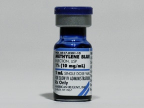
Why so blue? - Methylene blue in Distributive Shock

Clinical Scenario:
You are working in Trauma Critical Care when a middle aged male with end-stage liver disease presents with altered mental status.  He is hypotensive  and tachycardic.  You resuscitate him with IV fluids, start broad spectrum antibiotics and initiate vasopressors.  You confirm a source of infection with a paracentesis. Despite multiple and escalating doses of pressors, the patient's blood pressure continues to fall.  You perform a RUSH exam, run through your differential diagnosis again, and confirm that all lines and tubes are connected appropriately.  As you prepare his family for the worst and sign him out to the ICU, your colleague suggests - what about methylene blue?  Is it worth a shot?

Clinical Question:
Is there a role of methylene blue in management of septic shock?

Image source: webmd.com
Literature Review:
Methylene blue was initially developed as a dye for the textile industry, then used as a stain for TB, treatment for malaria, treatment of cyanide toxicity, and more recently methemoglobinemia.  Typical dosing for methemoglobinemia is 1-2mg/kg of 1% solution IV.

In a recent review performed by the Toxicology Department at UCSD published in the Journal of Emergency Medicine in 2013, the authors presented the evidence behind the utility of methylene blue in the septic patient [1].  

Methylene blue (MB) is useful in septic shock due to its ability to increase peripheral vascular resistance and its reversal of myocardial depression.  Its mechanism of action is thought to be due to inhibition of inducible nitric oxide synthase (up regulated by endotoxin and cytokines producing vasodilatory nitric oxide and guanylate cyclase (makes vasodilatory cGMP) [2,3].  There have only been two randomized controlled trials that studied MB in septic shock (n=20; n=30).  In these studies MB was found to increased mean arterial pressure (MAP) and decrease vasopressor requirements, but there was no statistically significant difference in survival rates.

Other studies reviewed for this publication were case series and case reports.  In these, varying doses of MB were used ranging from 1-3mg/kg over 10-20 minutes, which all increased MAP, systemic vascular resistance (SVR), and mean pulmonary arterial pressure.  However MAP and SVR returned to baseline 2-4 hours after MB administrations.

With doses greater than 4mg/kg or rapid or prolonged (greater than 6-10 hrs) infusion, potential side effects of MB use includes serotonin syndrome reaction (if already taking serotoninergic agents, MB inhibits monoamine oxidase), methemoglobinemia (if G6PD deficiency, or at high doses MB acts as oxidizer instead of reducer), platelet aggregation/reduction, and vasoconstrictive effects with possible decreases in splanchnic perfusion and arterial oxygenation (do not use in ARDS or pulmonary hypertension) [1,3,4].  The clinical effects of MB persist for 2-3 hours (half-life=102 minutes) and should be preferably administered centrally as peripheral administration can cause cutaneous necrosis [3,4].

Take home points:
-Methylene blue may prove a useful adjunct to vasopressors in cardiovascular collapse.  
-Remember additional shock management strategies include adequate volume resuscitation and source control, inotropy, respiratory support, correction of electrolytes (K, Ca, Mg, Phos) and glucose, consideration of endocrine dysfunction (thyroid, adrenal, vasopressin), and ruling out obstructive etiologies.

References:
1. Lo JC, Darracq MA, Clark RF. A review of methylene blue treatment for cardiovascular collapse. J Emerg Med. 2014 May;46(5):670-9.
2. Landry DW, Oliver JA. The pathogenesis of vasodilatory shock. N Engl J Med 2001; 345(8): 588-595.
3. Kirov MY, Evgenov OV, Evgenov NV, Egorina EM, Sovershaev MA, Sveinbjørnsson B, Nedashkovsky EV, Bjertnaes LJ. Infusion of methylene blue in human septic shock: a pilot, randomized, controlled study. Crit Care Med 2001; 29(10): 1860-1867.
4. Juffermans NP, Vervloet MG, Daemen-Gubbels CR, Binnekade JM, de Jong M, Groeneveld AB.. A dose-finding study of methylene blue to inhibit nitric oxide actions in the hemodynamics of human septic shock. Nitric Oxide 2010; 22(4): 275-280.

Submitted by Lydia Luangruangrong, PGY-3.
Edited by  Steven Hung (@DocHungER), PGY-2
Faculty reviewed by Chris Holthaus

Tuesday, November 18, 2014

Expert Commentary: Troubleshooting Hypoxia on the Vent

In emergency medicine education, we tend to focus on establishing the airway but spend less time discussing the aftercare and managing the ventilator.  While we have spent more time on this recently, thanks to Brian Fuller and his research on the importance of low tidal volume ventilation in the ER (see EM Journal club summary and podcast from February 2014),  most of us are still more comfortable putting the ET tube in than managing the vent.

Luckily for us, here at WashU we have some EM-Critical Care wise guys who can teach us a thing or two, and today we share with you Brian Fuller's method for trouble-shooting hypoxia on the vent, forwarded to us by PGY-3 Brendan Fitzpatrick:

>>From: Fitzpatrick, Brendan
>>To: Fuller, Brian
>>Subject: vent desats

Dr. Fuller,

Good working with you last night. I was trying to recall how you broke down desats on the vent last night, but somewhere between little sleep and my kids' halloween parade, I've lost the finer points.

In all your free time, would you mind jotting down what you told me so I can review it?

thanks,

Brendan


>From: "Fuller, Brian"
>To: "Fitzpatrick, Brendan"
>Subject: RE: vent desats


For the purposes of acute deterioration (in the form of hypoxia) on the ventilator, we are gonna talk about two airway pressures: peak pressure and plateau pressure. As an aside, mean airway pressure is the average pressure over one cycle of inspiration and expiration. It is largely governed by PEEP and I:E ratio. It really governs oxygenation- higher it is, more you open up stiff alveoli in sick vented patients.


Peak pressure is the summation of pressure generated from: 1) tidal volume and compliance; 2) resistance and peak inspiratory flow; and 3) PEEP
 

Plateau pressure is a reflection of compliance. Think "how stiff the lungs are"; or "how much transalveolar stretch is occurring".
Compliance is ∆ volume/∆pressure. Specifically, tidal volume/(plateau pressure - PEEP)


So the first thing I do when somebody becomes acutely hypoxic on the ventilator is to look at their peak airway pressure:

1. If decreased: you have an air leak or the patient is hyperventilating/tugging hard and therefore pulling the airway pressures down. Air leak would be something like: bronchopleural fistula, the chest tube you just put in has a leak in the system, your ETT has migrated or cuff has a leak and air is escaping.

2. If increased: see above- this could either be primarily a compliance or resistance problem. So your next step is to look at the plateau pressure to figure out where the problem lies.

If no change in plateau, you therefore have a bigger difference between the peak pressure and the plateau pressure than existed before the hypoxic event. See above for what governs these pressures, so you can tell that this is therefore a resistance problem. Think: airway obstruction from bronchospasm, clogging of the ETT with secretions, kinking of the ETT.


If plateau pressure is also increased, you now have a situation where the peak and plateau pressures both increased. See above for what governs these pressures, so you can tell that this is therefore a compliance problem. Think: pulmonary edema, abdominal distention, pneumothorax, atelectasis, etc.

3. If no change: think "Something made my patient hypoxic but didn't change my airway pressures." Not a lot of stuff does that. Think: pulmonary embolism, PFO.

Sometimes it is difficult to figure out "Is the ventilator and my ventilator settings the problem, or is this a patient problem?" If you remove the patient from the ventilator, and therefore take that out of the equation, bag them and they get better, it is probably a ventilator problem. If you bag them and they stay bad, it is probably a patient problem.

Hope this helps. Hit me back with questions PRN.

Feel free to share with others.

Be good man.
B


For those of you who like pictures, here is a visual representation of the algorithm:

Want to do a little more reading or watching about this? Life in the Fast Lane had a nice review of Pulmonary Mechanics.  I recommend the second Eric Strong video on pulmonary pressures as a supplement to understanding the above material (and hell, it only takes 9 minutes to watch).

Expert Commentary by Brian Fuller
Visual aids by Maia Dorsett (@maiadorsett), PGY-3 
Expert inquiry, sharing skills and parenting by Brendan Fitzpatrick, PGY-3.